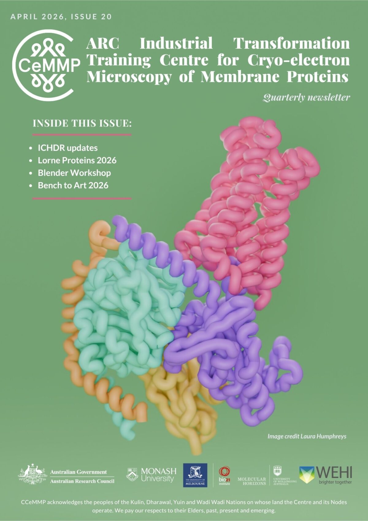
Cryo-electron Microscopy of Membrane Proteins
What are membrane proteins?
Cells are the basic units of life. Cells may have individual roles, such as those of the immune system that circulate in our bodies and respond to foreign invasion, but more commonly are part of larger aggregates that make up our vital organs. Cells are separated from each other and the environment by membranes, a barrier that is comprised of a lipid bilayer with embedded or associated proteins.
These membrane proteins allow the cell to sense their external environment and can control the internal cell environment through regulation of the movement of substances in and out of the cell, or by changing the activity within the cell. A major class of membrane proteins are termed receptors, and these receptors are the conduits of intracellular communication and allow the coordinated control of cells that underpins complex physiology. Disruption of normal cellular function leads to disease and altered behaviour of membrane proteins can contribute to disease, but targeted regulation of these proteins can also lead to disease resolution.
As such membrane proteins are the largest class of therapeutic drug targets.
How do we visualise membrane proteins?
To visualise membrane proteins or parts of membrane proteins we use a method called, cryo-electron microscopy (cryo-EM). In EM, a coherent beam of electrons replaces the light source (photons) of conventional microscopes. The properties of the electron beam enable much higher resolutions to be achieved. Cryo-EM is a specialised form of EM where samples are frozen in a non-crystalline form of ice, termed vitreous ice, which allows images of samples captured in their native state to be visualised. Cryo-EM can be applied to purified proteins that are frozen in very thin vitreous ice so that only a single protein is present in any section of the ice generating an atomic level 2D picture of that protein (single particle analysis, SPA).
However, during freezing the protein will have multiple different orientations with each of these leading to 2D images of the protein at different angles. By capturing images of 100s, 1,000s or even 1,000,000s of particles, sophisticated software allows us to reconstruct these 2D images into a high-resolution 3D map of our protein of interest that we use to determine the structure of the protein. The advances in vitrification, direct electron detectors, and the sophisticated software that collectively allow cryo-EM to now be used for protein structure determination formed the basis of award of the 2017 Nobel Prize in Chemistry.
Why use Cryo-EM to visualise Membrane Proteins?
Membrane proteins, by their nature, are often highly dynamic and unstable if removed from their native lipid environment. This has made the structure of membrane proteins very difficult to be solved by other methods x-ray crystallography or NMR. In contrast, cryo-EM can be applied to very small amounts of purified protein, it does not require the proteins to form stable crystals for imaging, and it can allow high-resolution reconstruction of dynamic proteins.
Cryo-EM has revolutionised our ability to determine 3D membrane protein structure and has opened up new potential for structure-assisted drug discovery and development.
These membrane proteins allow the cell to sense their external environment and can control the internal cell environment through regulation of the movement of substances in and out of the cell, or by changing the activity within the cell. A major class of membrane proteins are termed receptors, and these receptors are the conduits of intracellular communication and allow the coordinated control of cells that underpins complex physiology. Disruption of normal cellular function leads to disease and altered behaviour of membrane proteins can contribute to disease, but targeted regulation of these proteins can also lead to disease resolution.
As such membrane proteins are the largest class of therapeutic drug targets.
Recent News